* Our Advanced Anatomy content is developed for High School and/or Introductory College Level Students.
It is estimated that there are 60,000 miles of blood vessels in the human body made up of arteries, veins and capillaries. Their function is to deliver oxygen (O2) and nutrients to all the tissues of the body via arteries and take away the carbon dioxide (CO2) and waste via veins.
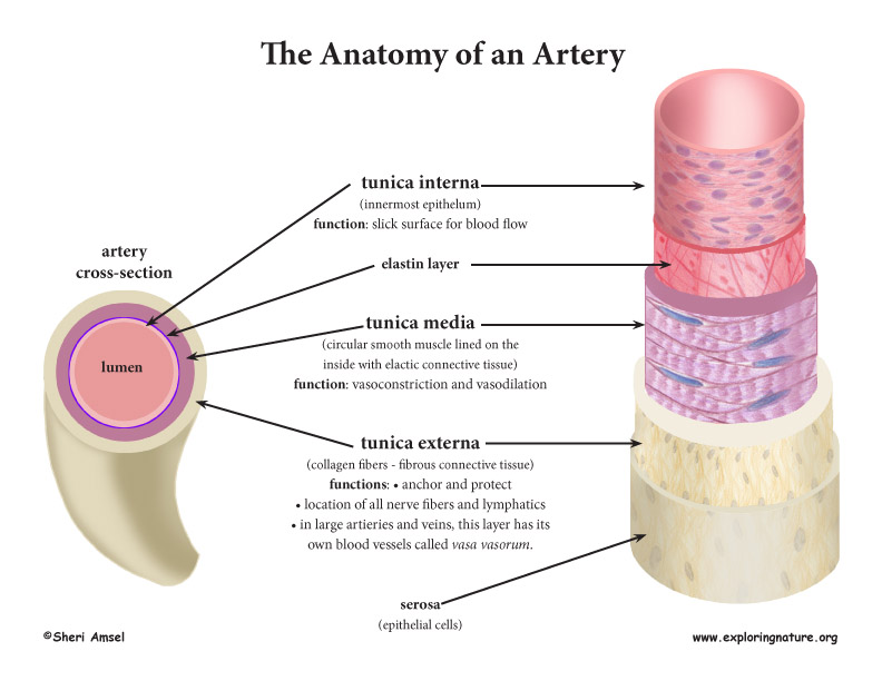
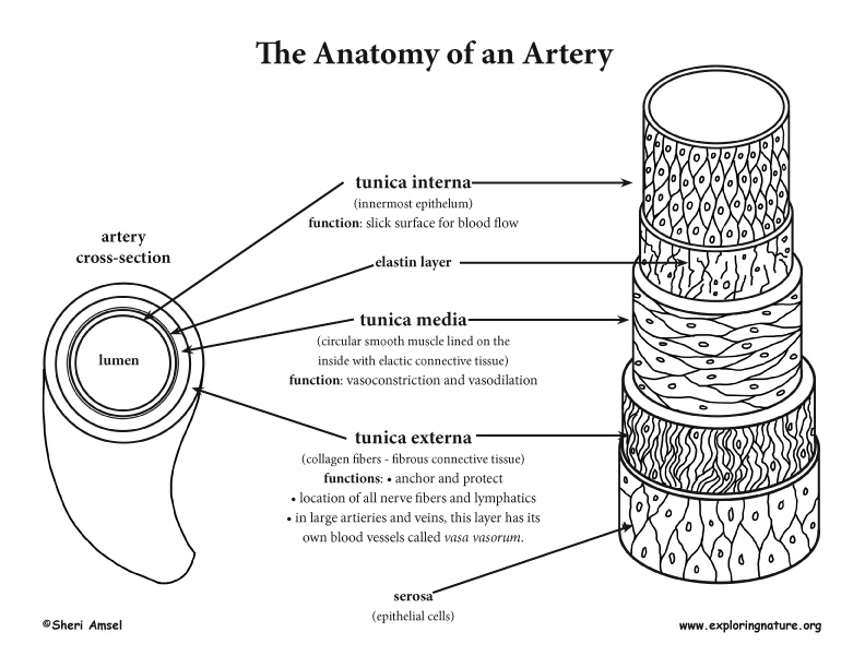
Blood Vessel Structure
The structure of the blood vessels is designed for the ebb and flow of blood as its pumped through the body. Both arteries and veins (not capillaries) are made up of several layers or tunics.
1. The innermost layer is called the tunica interna. It is made up of endothelium and functions as a slick surface for unhindered blood flow.
2. The next layer is the tunica media. It is made up of smooth muscle with a very thin layer of elastin lining its inside. The tunica media has vasomotor fibers from the sympathetic nervous system that regulate blood flow by signaling for this layer to vasodilate or vasocontrict as needed.
3. The outside layer is the tunica externa. It is made up of collagen fibers that anchor and protect the blood vessel. There are nerve fibers and lymphatics in this layer and in the largest blood vessels (aorta and vena cavas) there are even blood vessels feeding it oxygen called vasa vasorum.
Arteries
Arteries generally carry blood away from the heart. There are three types:
1. Elastic Arteries - These are conducting arteries. They are the largest, most thick walled, most elastic and close to the heart (aorta and main branches). They have an elastic lamina sandwiching the tunica media to protect it from the changes in blood pressure from the pumping heart. They do not do much vasoconstriction.
2. Muscular Arteries - These are distributing arteries. They are medium to smaller arteries that conduct blood to different parts of the body.
They have more smooth muscle in the tunica media, so are active vasoconstrictors.
3. Arterioles - They are the smallest arteries. They feed the capillary beds. The vasoconstriction and vasodilation of the arterioles are the most important factor in determining blood flow in the capillary bed.
Test your knowlege of Blood Vessel Anatomy with this Labeling Page.
Vein Structure
Vein structure is similar, but not identical, to arteries. They are made up of the same layers or tunics but they differ in the following ways:
• The vein lumen is larger.
• The walls of the tunics are thinner with little smooth muscle.
• The tunica externa is larger than the tunica media.
• The veins contain the blood reservoirs of the body. At any one time, the veins of the body contain 65% of the body’s blood. This is why the veins are considered to be capacitance vessels.
• Veins have lower blood pressure than arteries. They are further from, so less influenced by, the pumping action of the heart.
• Valves in the veins prevent blood back flow in their low pressure environment. There are more valves in the limbs where the blood is flowing against gravity. The valves are made up of a flap of the tunica interna. Their shape is like the semilunar valves of the heart.
• Breathing in and out (respiration) helps to pump venous blood back to the heart, as does muscular movement.
*The London Guards, who stand very still all day, are taught to flex their muscles as they stand to keep their venous blood flowing back to the heart.
• Varicose veins are the result of leaky valves. The veins become, over time, dilated and tortuous. Leaky valves result from heredity, obesity, pregnancy and standing professions.
• Hemorrhoids result from raised venous pressure from straining during defecation or childbirth. They are essentially varicose veins of the anal canal.
Venous Sinuses
The venous sinuses are specialized, flattened veins with thin endothelium walls. They are supported by the surrounding tissues.
1) There are venous sinuses in the cranium - intracranial venous sinuses (dural sinuses) that drain the blood from the brain and the cerebrospinal fluid around the brain. It is endothelium underlined by the dura mater (the brain covering).
2) There is a venous sinus around the heart - coronary sinus that drains the myocardium of the heart.
Capillaries
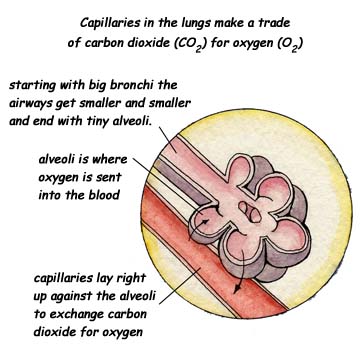
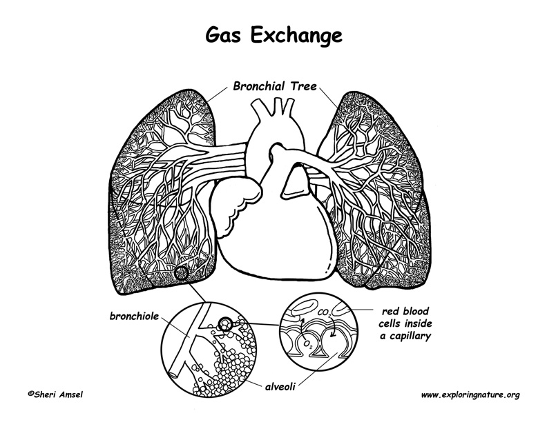
Function: Capillaries are the smallest blood vessels in the body.Some capillaries are so small, they are just one cell thick. This makes them well suited for exchanging gases and nutrients between the blood and the body's organs and tissues. In the lungs, oxygen is passed from the capillaries into the alveoli, while carbon dioxide is passed out from the alveoli to the capillaries for disposal. In the small intestine, food nutrients, like glucose, pass from the small intestine into the capillaries. From there, they deliver the nutrients to every part of the body that needs it.
Structure: Capillaries are a dense network, branching throughout all the body’s tissues. They lay right up against organs and tissues for easy exchange of food and oxygen for wastes. They are very thin-walled with just a tunica interna. They have no muscle or connective tissue. Most tissues have a rich capillary supply - EXCEPT tendons, ligaments, cartilage, the cornea and the lens of the eye.
Capillaries are continuous or fenestrated:
1. Continuous Capillaries:
• are abundant in the skin and muscles.
• have an uninterrupted lining with tight junctions.
• make up the blood-brain barrier.
• some have some transport - pinocytosis
2. Fenestrated Capillaries:
• are made up of endothelial cell with gap junctions - are fenestrated.
• are found in areas of active absorption - small intestine, kidneys, etc.
• are also found where secretion occurs - endocrine glands, glomerular capillary beds, etc.
Capillary Beds
There are 2 types of Capillary Beds:
1. Vascular Shunt (metarteriole or thoroughfare channels) - short vessels connecting artioles and venules.
2. True Capillaries:
• Branch off thoroughfare channels or artioles
• Take part in gas and/or nutrient exchange with tissue cells
• Blood goes though true capillaries in regions of the body most needing them - i.e. the viscera after eating, the skeletal muscles after exercising, etc.
Sinusoids
• Modified capillaries that connect arterioles and venules in the liver, lymphatics and some endocrine glands.
• Often lined with phagocyte cells (spleen)
• Usually tortuous, flowing slowly to be “processed” (in the liver this is done by Kupffer Cells).