Your skin, which is also called integument or epithelium, is an organ. It is actually the largest organ of the body making up about 7% of your body weight. That means that a 200-pound adult has about 3,000 square inches of skin, which weighs about 14 pounds.
The function of the skin is:
1. Maintains body temperature.
2. Protects against abrasions, microorganisms, dehydration and UV light.
3. Excretes waste (water, salts, etc.)
4. Absorbs sunlight and synthesizes Vitamin D.
5. Receives stimuli - touch, pressure, heat, and pain.
6. Provides immunity.
The skin has three layers made up of more than a half million cells.
l. Epidermis
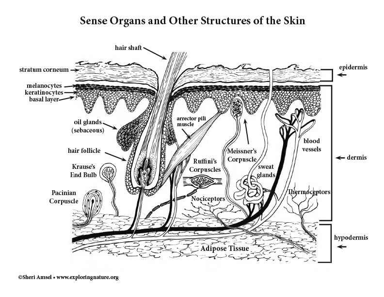
The outer layer of the skin is called the epidermis, which is made up of several layers and has constant cell regerneration. The epidermis has cells called melanocytes. They make melanin, a pigment that protects us from ultraviolet (UV) radiation, the dangerous part of sunlight.
5 Layers of the epidermis are:
1. Stratum corneum (outermost layer) - many layers of hard, dead cells that are water repellent and made of keratin fibers (avascular). These cells protect us by shedding when we touch or run up against another surface. They also keep foreign bodies out and our fluids in. This layer makes the skin waterproof.
2. Stratum lucidium - found in areas of skin with the greatest thickness (palms and soles of feet)
3. Stratum granulosum - where keritinization begins. This layer is made up of the living cells (keratinocytes) that become the stratum corneum.
4. Stratum spinosum - layer with langerhan's cell and gives skin stength and flexibilty
5. Startum basale (Stratum germinativum) - all layers of epidermis arise from this layer, including the vital keratinocytes.
ll. Dermis
The skin’s second layer is called the dermis. Layers of the dermis are:
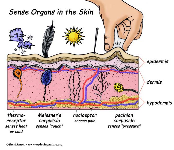
1. Papillary Layer - superficial layer (made of loose areolar connective tissue) gives rise to dermal papillae that reach up through the epidermis and aid in gripping. Also where Meissner's corpuscles are located for detecting touch.
2. Reticular Layer deepest layer of the dermid (give strength to skin) - where the following structures are located:
• Blood vessels - The blood vessels in the face’s dermis will expand in cold weather allowing more warm blood to flow there (and keep your exposed face warm). This is why your face gets red in cold weather. This is also what happens when you blush.
• Sense organs - respond to stimuli like heat, cold, pressure, touch and pain. They receive the information and pass it onto the brain for action.
• Oil and sweat glands -
• Hair follicles - The many tiny hairs on your skin are rooted in the dermis. They are attached to the smallest muscles of the body called arrector pili muscle. Cold air or a sudden fright makes these tiny muscles contract and pull the small hairs upright. This is what gives you goosebumps. Hair follicles extend down into the dermis or even the hypodermis.
lll. Hypodermis
The deepest layer of the skin is the subcutaneous layer or the hypodermis. The hypodermis is a layer of protective fibers and fat. About half of all your body’s fat is found in the hypodermis.
The function of the hypodermis:
1. Fat and blood vessels help maintain body temperature.
2. Fat cushions the body from trauma
3. Attaches skin to body
Fingerprints are friction ridge patterns on our finger and toe pads that help us grip things without slipping. No two people have the same fingerprints.
The skin reacts to the external environment, but also to what is going on inside the body. A fever (felt through the skin) tells us there is an infection that the body is fighting off. A rash shows an allergic reaction of some kind. Wrinkles show aging and freckles can mean sun damage.
The most common form of cancer in the U.S is skin cancer. Skin cancer can be prevented by covering or applying sunscreen to the epidermis.
Skin heals from injury all time. Tiny scrapes to the outer layer of epidermis triggers new cells (keratinocytes) to grow to replace the ones that are scraped off. Injuries that reach down into the dermis (or deeper) where the blood vessels are located will cause bleeding. Sometimes the area heals leaving no trace, but sometimes connective tissue fibers will replace some of the skin cells leaving a scar.
When you research information you must cite the reference. Citing for websites is different from citing from books, magazines and periodicals. The style of citing shown here is from the MLA Style Citations (Modern Language Association).
When citing a WEBSITE the general format is as follows.
Author Last Name, First Name(s). "Title: Subtitle of Part of Web Page, if appropriate." Title: Subtitle: Section of Page if appropriate. Sponsoring/Publishing Agency, If Given. Additional significant descriptive information. Date of Electronic Publication or other Date, such as Last Updated. Day Month Year of access < URL >.
Amsel, Sheri. "Integumentary System Overview" Exploring Nature Educational Resource ©2005-2025. March 20, 2025
< http://www.exploringnature.org/db/view/Integumentary-System-Overview >