The Peripheral Nerves of the Neck, Shoulders and Arms
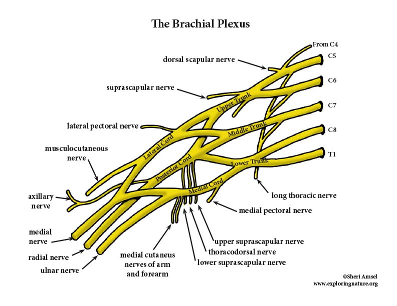
Many spinal nerves form interlacing nerve networks – the cervical, brachial, lumbar and sacral nerve plexuses. The cervical plexus in the neck involves the upper 4 cervical nerves and forms the phrenic nerve which supplies both sensory and motor innervation to the diaphragm which is important for breathing. The brachial plexus in the neck and armpit (axilla) involves C4-T2 and innervates the upper limb (arm and shoulder). These give rise to the axillary nerve, musculocutaneous nerve, median nerve, ulnar nerve and radial nerve (and a few others).
The axillary nerve comes out behind the head of humerus to innervate the shoulder joint and the deltoid and teres minor muscles.
The musculocutaneous nerve comes down the anterior arm innervates flexion of theforearm and sensory for the laterally forearm.
The median nerve comes all the way down the central anterior arm to innervate the forearm flexor muscles and activate pronation of the forearm, flexion of the wrist and fingers and opposition of the thumb. It receives sensory from the forearm skin and the lateral palm.
The ulnar nerve comes down the medial side of the arm, goes behind medial condyle of the elbow, down the medial forearm and innervates muscles of the forearm and fingers for wrist and medial finger flexion, adduction and abduction. It receives sensory from the medial side of the hand.
The radial nerve is the largest of the nerves off the brachial plexus. It comes down the posterior arm, then wraps around the anterior aide of the lateral epicondyle of the elbow, then splits into a superficial branch and deep branch. It innervates all the extensor muscles of the upper limb and receives sensory from the skin of the posterior arm.
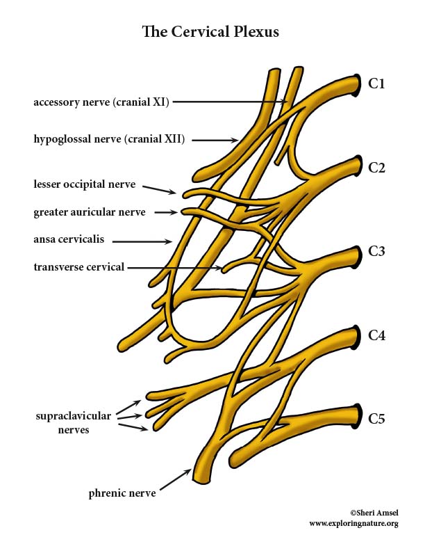
The Cervical Plexus
The cervical plexus is deep in the neck and is fed by the ventral rami of C1-C4. It supplies both sensory (afferent) and motor (efferent) fibers to the phrenic nerve which controls the diaphragm - the main muscle that allows breathing movements. The cervical plexus also brings in sensory impulses from the skin of the neck, ears and shoulders. There is some motor for the muscles of the anterior neck as well.
The Brachial Plexus
The brachial plexus is the source of almost all the nerves that innervate the upper limb (arm and shoulder. It lies in the neck and armpit (axilla) and involves nerve fibers from C4 -T2. These give rise to the axillary nerve, musculocutaneous nerve, median nerve, ulnar nerve and radial nerve (and a few others).
The Lumbosacral Plexus (The Peripheral Nerves of the Pelvis, Buttock and Lower Limbs)
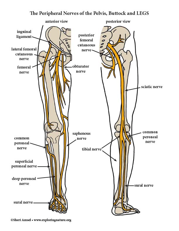
The lumbosacral plexus is the overlapping of the lumbar and sacral plexuses and innervates the lower limb and parts of the abdomen, pelvis and buttock.
The lumbar plexus comes off L1-L4. It innervates the anterior and medial thigh and parts of the abdominal wall and iliopsoas muscle. It’s largest branch, the femoral nerve, splits into many branches to innervate the anterior thigh muscles for thigh flexion and knee extension. It receives sensory input from the anterior thigh and medial leg between the knee and foot. The obturator nerve innervates the medial thigh adductor muscles.
The sacral plexus comes off L4-S4. It’s largest branch is the sciatic nerve, which is made up of the tibial nerve and the common peroneal nerve. The sciatic nerve enters the back of the thigh just medial to the hip joint and gives off branches to innervate the hamstring muscles, which are thigh extensors and knee flexors. Right above the knee it splits into the tibial and common peroneal nerves.
The tibial nerve goes behind the knee (called the popliteal fossa) and innervates the posterior calf muscles. It receives sensory from the skin behind the calf and the sole of the foot. It gives off the sural nerve which receives sensory from the posterior-lateral calf and the plantar nerves which innervates the foot.
When the common peroneal nerve splits off the sciatic nerve, it wraps around the head of the fibula and splits into the superficial and deep branches which innervate the anterior-lateral calf muscles, which are extensors and dorsiflexors of the foot. It receives sensory from the knee joint, lateral calf and top (dorsum) of the foot.
The sacral plexus also gives off the superior and inferior gluteal nerves which innervate the gluteal muscles of the buttock and a lateral muscle of the thigh (tensor fascia lata). It also gives of the pudendal nerve, which innervates structures in the pelvis.
The Lumbar Plexus
The lumbar plexus comes off L1-L4. It innervates the anterior and medial thigh and parts of the abdominal wall and iliopsoas muscle. It’s largest branch, the femoral nerve, splits into many branches to innervate the anterior thigh muscles for thigh flexion and knee extension. It receives sensory input from the anterior thigh and medial leg between the knee and foot. The obturator nerve innervates the medial thigh adductor muscles.
The Sacral Plexus
The sacral plexus comes off L4-S4. It’s largest branch is the sciatic nerve, which is made up of the tibial nerve and the common peroneal nerve. The sciatic nerve enters the back of the thigh just medial to the hip joint and gives off branches to innervate the hamstring muscles, which are thigh extensors and knee flexors. Right above the knee it splits into the tibial and common peroneal nerves.
The tibial nerve goes behind the knee (called the popliteal fossa) and innervates the posterior calf muscles. It receives sensory from the skin behind the calf and the sole of the foot. It gives off the sural nerve which receives sensory from the posterior-lateral calf and the plantar nerves which innervates the foot.
When the common peroneal nerve splits off the sciatic nerve, it wraps around the head of the fibula and splits into the superficial and deep branches which innervate the anterior-lateral calf muscles, which are extensors and dorsiflexors of the foot. It receives sensory from the knee joint, lateral calf and top (dorsum) of the foot.
The sacral plexus also gives off the superior and inferior gluteal nerves which innervate the gluteal muscles of the buttock and a lateral muscle of the thigh (tensor fascia lata). It also gives of the pudendal nerve, which innervates structures in the pelvis.
When you research information you must cite the reference. Citing for websites is different from citing from books, magazines and periodicals. The style of citing shown here is from the MLA Style Citations (Modern Language Association).
When citing a WEBSITE the general format is as follows.
Author Last Name, First Name(s). "Title: Subtitle of Part of Web Page, if appropriate." Title: Subtitle: Section of Page if appropriate. Sponsoring/Publishing Agency, If Given. Additional significant descriptive information. Date of Electronic Publication or other Date, such as Last Updated. Day Month Year of access < URL >.
Amsel, Sheri. "Peripheral Nervous System - Nerves (Advanced)" Exploring Nature Educational Resource ©2005-2024. December 13, 2024
< http://www.exploringnature.org/db/view/Peripheral-Nervous-System-Nerves-Advanced >